Make informed decisions for your operation with information delivered right to your inbox.
Get the latest tools, innovations and science-based information for the Canadian beef industry, including seasonal production considerations and economic analyses.
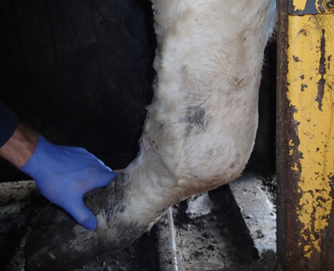
Lameness occurs when an animal has leg or foot pain that affects how they move. Lameness is an animal health and welfare concern, as well as a production issue. Pain due to lameness often limits growth because animals may be reluctant to eat or drink.
There are many types of lameness, with several different causes, many of which are inter-related. Lameness can be caused by genetics, environment, nutrition, injury, or infection.
Key Points
Lameness in beef cattle is a painful condition and a concern for animal health and welfare. Lame cattle often have a lower rate of gain which also makes lameness a production and economic issue.
Unhealthy cattle are at a greater risk for developing lameness and lameness is also a risk factor for other diseases. For example, lameness in cattle may be associated with Bovine Respiratory Disease (BRD).
Prior to transport, carefully consider whether animals are suited for shipping. Market cows are at a greater risk for lameness and injury during transport.
Lameness can be caused by infection, injury, nutrition, genetics, or a combination of these factors, sometimes making it a challenge to properly diagnose.
Not all lameness is caused by foot rot and an accurate diagnosis is important for treatment success as well as to avoid unnecessary treatment with antibiotics. If an animal is thought to have foot rot but does not respond to treatment, foot rot was likely a misdiagnosis.
Nutritional-related lameness, including laminitis (founder) and mycotoxin-caused necrosis, may be prevented by careful feed bunk management and feed testing.
Injuries from abrasive flooring or muddy pen conditions can provide an entry point for other infections.
Injuries, such as frostbite, sandcracks, breaks, or sprains, may not be treatable. Alternate options, such as pain management or euthanasia, should be assessed in consultation with a veterinarian.
There are several risk factors that can predispose some cattle to lameness including: muddy and contaminated pen environments; facility design flaws such as abrasive or slippery flooring; the presence of sharp protrusions or objects; improper animal handling; agitated cattle behaviour; other diseases such as Mycoplasma bovis; and heavy cattle carrying a lot of weight.
Some management practices may help to reduce the risk of lameness, including regularly cleaning pens; disinfecting hoof-trimming tools; removing sharp objects and protrusions from pens and handling areas; vaccinating/preconditioning cattle to strengthen immunity; practicing low-stress animal handling; and using proper facility and flooring design.
Importance to the Beef Industry
Lameness in beef cattle is an animal health and welfare issue that also has production and economic consequences for cow-calf and feedlot operators. Studies show that lame feedlot cattle grow more slowly than cattle that are not lame and steers diagnosed with foot rot during the finishing period need up to two weeks longer to reach slaughter weight. A lower rate of gain can be costly in terms of welfare, labour, pen space and feed.
Many animals will recover with appropriate treatment. Chronic cases may be salvaged, provided all pharmaceutical withdrawal requirements have been met and the animal can be transported without additional suffering. Humane euthanasia on the farm is recommended when animal welfare is severely compromised and not expected to improve.
Animal showing signs of lameness (Photo credit: Eugene Janzen and his team).
Incidence
In a study of 28 western Canadian feedlots over 4 years, researchers found that hoof related lameness accounted for 26% of all treatments that were administered to animals. Of those treated for hoof related lameness, the most common causes were foot rot (90%), followed by digital dermatitis (8%) and toe tip necrosis (2%). Foot rot occurred throughout the feeding period at a relatively steady rate whereas digital dermatitis was more common between 80-190 days on feed and toe tip necrosis was most common during the first 100 days on feed.
Cattle sourced from backgrounding and grass-backgrounding operations had a higher risk of developing hoof related lameness than cattle sourced from ranches or auction marts. Calves were higher risk than yearlings, and cattle from mall feedlots were higher risk than those in large feedlots.
Lameness is more common in unhealthy cattle and is also a risk factor for developing additional diseases. In an older western Canadian feedlot study, cattle diagnosed with lameness due to injury, joint infection, or lameness with no visible swelling were associated with a diagnosis of Bovine Respiratory Disease (BRD). In another study, researchers found that lameness accounted for 37.4% of cattle in the chronic illness pen, with another 10.9% of cattle being diagnosed with both respiratory disease and lameness.
Transport practices can affect lameness and may make any lameness issues worse. However, the risk is reduced when cattle are healthy and fit at the time of loading. A 2007-08 survey of 50,000 beef cattle transported in Ontario identified 79 lame cattle. A 2008 survey that looked at more than 290,866 beef cattle transported in Western Canada found 37 lame cattle, however the study also reported that lameness was considerably more common in market cows than in fed cattle, feeders or calves. As well, the survey found that the likelihood of lameness increased with the duration of transport.
Causes & Types of Lameness
Four common causes of lameness include:
Infection (i.e. foot rot, digital dermatitis, toe tip necrosis, infectious arthritis)
Emerging causes of lameness, including digital dermatitis and toe tip necrosis, are becoming better understood, as are the multiple inter-related causes of lameness. For example, when nervous cattle (which may be attributed to genetics) scramble on hard flooring, they may damage the sole of the foot (injury), allowing bacteria to enter and colonize the foot (infection). Each of these types of lameness are associated with specific risk factors. Trauma or wet conditions that affect skin or hoof integrity can cause lameness directly and also provide an avenue for bacteria to enter and colonize a wound.
Producers should not assume that lame cattle have foot rot without close observation, to avoid unnecessary administration of antibiotics.
Foot Rot
Foot rot is a painful condition that causes lameness and can affect any class of cattle, whether in a feedlot, corral, or pasture. Foot rot is often characterized by a sudden onset of lameness and is worse during wet conditions. People sometimes assume any lameness is caused by foot rot however this is not true.
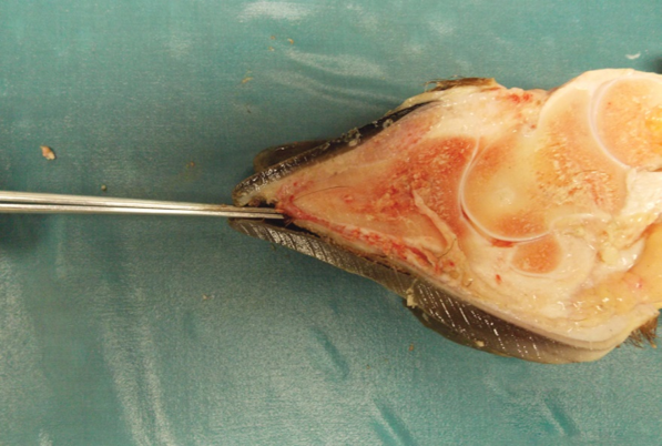
Foot rot is highly infectious and is caused by bacteria, most commonly Fusobacterium necrophorum. The infection originates in between the claws of the hoof and may be characterized by heat and swelling in between the claws, as well as along the coronary band where the hoof meets the skin. If it is not identified and treated promptly, the infection can move elsewhere into bones, joints, or tendons, causing delayed recovery or other complications. Fortunately, foot rot infections almost always respond well to treatment.
Dr. Jacques van Zyl is an Ontario veterinarian that works with beef cattle. He says adding a pain control product to a treatment protocol for diseases such as foot rot is manageable. Learn more.(Photo courtesy of Metzger Veterinary Services)
When breeding bulls are afflicted with foot rot, there can be negative long-term consequences for the entire herd. The increase in body temperature, stress, and pain caused by foot rot can reduce sperm production for a period of time, as well as reduce a bull’s libido, leading to open cows. Talk to your veterinarian to determine if vaccinating against foot rot infections is a useful prevention method.
Foot rot infection demonstrating affected tissue between the claws of the hoof. (Phooto credit: Eugene Janzen and his team).
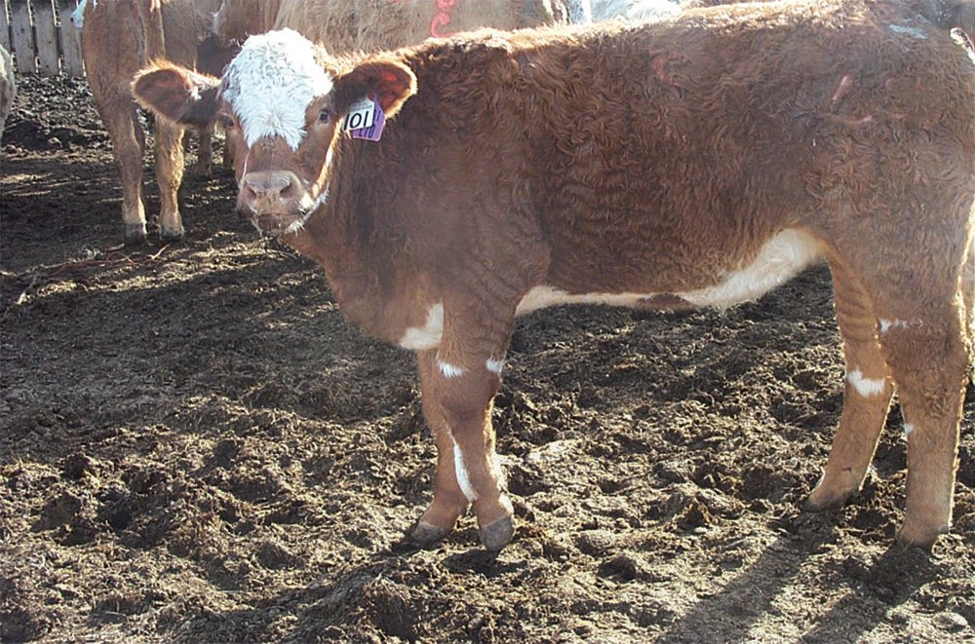
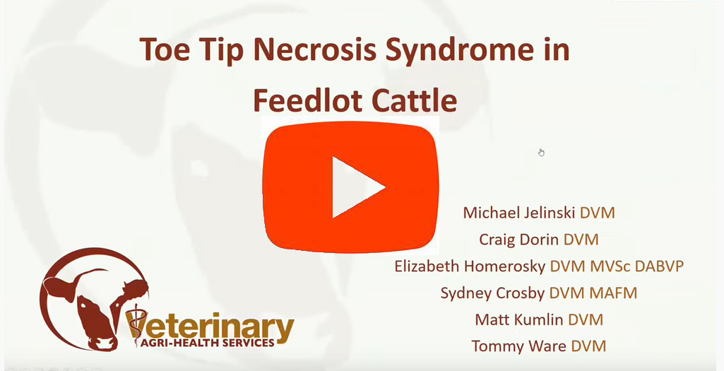
Toe Tip Necrosis
Toe tip necrosis is a lameness condition affecting the hind feet of feedlot cattle. It develops early in the feeding period, and often occurs within clusters of animals or cohorts. Sometimes referred to as a toe abscess or ulcer, apical white line disease, or P3 necrosis, it has been associated with improper processing or handling, abrasive flooring, as well as flighty animal behaviour. Careful, low-stress handling and proper flooring may help prevent the development of toe tip necrosis.
The symptoms of toe tip necrosis (hoof damage and fever) can be similar to injury, making it challenging to obtain a definitive diagnosis. It is a very painful condition discussed in a recent BCRC webinar (skip ahead to 41:20).
Many animals respond to antibiotics but high mortality rates are common with those that do not respond to medication. Tipping the tow to allow for drainage appears to help in some cases.
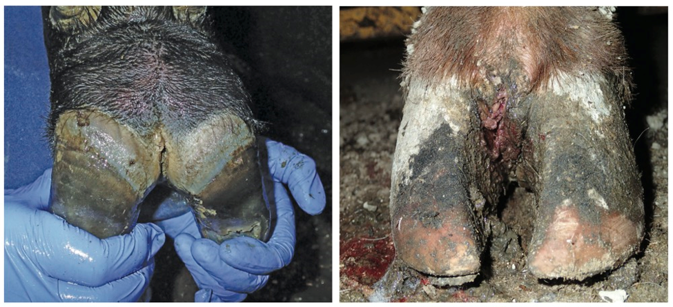
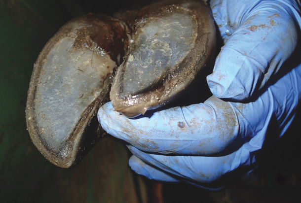
Research has found that the disease seems to occur from the outside in. The disease starts when the white line between the sole and the hoof wall separates, allowing bacteria to penetrate into the foot reaching into the corium, which is a very sensitive tissue. Inflammation of the corium causes pain, which is made worse if the infection continues into the foot and infects the P3 (coffin) bone.
Pressure builds as an abscess forms in the toes of a hind foot when toe tip necrosis infection is present. (Photo credit: Murray Jelinski). A cross-section of the hoof demonstrates separation between the hoof wall and the white line in an animal affected by toe tip necrosis. (Photo: Murray Jelinski).
Digital Dermatitis
Digital dermatitis, also known as hairy heel wart or strawberry foot rot, is a skin infection that affects the area around the dewclaws, between the claws, and sometimes the heel of the hooves. The disease is characterized by raised lesions often located between the claws and dewclaws. These lesions are very painful, may appear red and bleed easily if disturbed, and some may have long fibrous hairs. Animals appear to walk on their tip toes to avoid putting pressure on their heels.
Digital dermatitis is highly infectious but doesn’t respond well to injectable antibiotic treatment. Once an animal is infected with Treponema, the organism primarily associated with digital dermatitis, the animal will have it for life. To complicate things, digital dermatitis may occur simultaneously with foot rot. Footbaths can be an effective treatment however there are logistical challenges when trying to use footbaths to treat large numbers of animals.
It was long thought that digital dermatitis was only a dairy cattle disease, however it is being increasingly diagnosed in confined beef cattle. A recent webinar discussed digital dermatitis in Ontario, where veterinarians are seeing the disease appear. This seems to be more common in pack barns compared to slatted barns (skip to 26:53).
Joint Infections and Arthritis
An animal that has been sick over a longer period of time runs the risk of having the infection spread into bones or joints, making it very difficult to treat. For example, Mycoplasma bovis is commonly implicated in cases of pneumonia and mastitis. M. bovis can also travel through the animal’s bloodstream, sometimes settling in the ankle, stifle, hock, or elbow joints, leading to painful swelling and arthritis.
Histophilus somni is another organism that has been linked to arthritis. Unlike M bovis, there is a vaccine available for H somni.
Animals with joint infections may become chronically ill if they aren’t caught and treated in time, or if tissue damage compromises treatment. Pain management, and in severe cases, euthanasia may be required. Preventing the disease and learning to recognize these animals early on is the best strategy.
A swollen joint can cause lameness and become a serious health and welfare problem.
Laminitis (Founder)
Laminitis is a condition where the lamellae, the layer of tissue between the hoof wall and the coffin bone, is weakened or becomes separated. Laminitis is linked to rumen acidosis which is when the rumen pH drops causing rumen bacteria to produce toxins that can pass through the rumen wall. These toxins can cause swelling in the blood vessels of the hooves, leading to founder or laminitis. Laminitis is usually attributed to diets that are high in fermentable carbohydrates such as grain, and it may occur after a sudden or rapid change in rations.
Laminitis may be more common in cattle that are held on feed too long and there is no practical treatment once an animal is afflicted, other than ensuring animals are kept on soft footing in pens that are well bedded.
Hoof elongation and curled claws can sometimes accompany severe cases of laminitis or founder.
Mycotoxin-related Necrosis
Mycotoxins in feed caused by ergot bodies or other fungi during wet growing conditions can lead to lameness. Ergot restricts blood flow to the hooves and other extremities which can cause severe lameness and even hoof-sloughing or other debilitating health and welfare issues. These animals often become chronically ill and require pain management or euthanasia. Prevent mycotoxin-related necrosis by feed testing to avoid or dilute mycotoxin-contaminated feeds. If caught early, this condition can be reversed by removing the contaminated feed source from the ration.
Physical Injuries
Beef cattle may sustain physical injuries, including sprains, breaks, sand cracks, or frostbite. Injuries should be carefully examined to ensure the lameness isn’t caused by an infection. Treatment will depend on the extent of the injury and the course of action should be decided upon in consultation with your veterinarian.
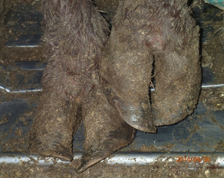
Sand cracks, which are vertical cracks in the hoof wall, are relatively common in range cattle in the Prairie Provinces. Sand cracks typically heal themselves through thickening of the hoof wall. Environment, exposure to moisture, or heavy animals may be contributing factors to the incidence of sand cracks. Provided the animal isn’t lame, treatment is often ineffective and unnecessary. If the animal does become lame, further inspection is warranted to treat for possible abscesses or infections.
Frostbite most commonly occurs in young animals although older cattle are not immune. When tissue freezes, blood circulation to the hooves is restricted, and tissue damage – either temporary or permanent – occurs. If the animal recovers from temporary frostbite damage, they can maintain a normal life, but permanent blood vessel damage occurs at a young age and hooves will not grow properly. In these cases, quality of life and production is compromised and euthanasia should be considered.
Genetic Problems
While management and environmental conditions can cause lameness issues, poor foot and leg conformation can also be a result of genetics. Genetic causes of lameness can be passed down to offspring and are sometimes slow to appear. Producers should work with veterinarians who can score herd bulls during breeding soundness evaluations to determine if there are structural issues. Producers should also be monitoring for foot and leg conformation in replacement females. Because lameness is multi-faceted, cattle that have proper foot and leg conformation will be better suited to withstand poor pen conditions or other non-infectious risk factors for lameness.
Management, Risks & Prevention
Accurate diagnosis is important for successful treatment and prevention of lameness. Veterinarians and researchers are beginning to learn more about common types of lameness including infectious causes such as toe tip necrosis and digital dermatitis. As new information emerges, producers and feedlot staff are learning how to identify and more accurately diagnose the differences between lameness diseases. Producers should not assume that all lame cattle have foot rot without close observation, to avoid unnecessary administration of antibiotics.
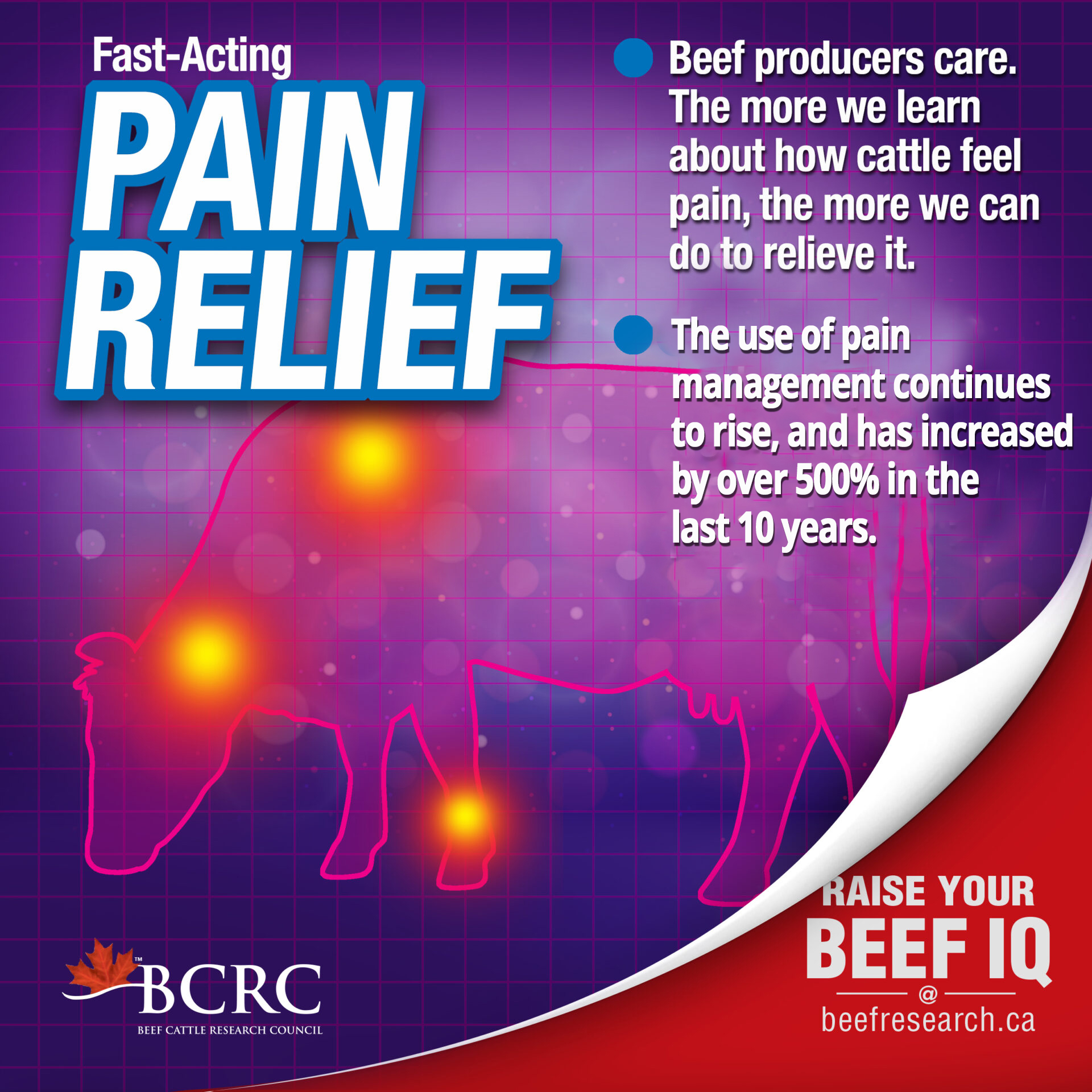
Pain management is an important consideration to improve both welfare and production outcomes in a lame animal. Some veterinarians and producers may use steroids, such as dexamethasone, as an anti-inflammatory in certain cases. The increased availability of non-steroidal anti-inflammatories (NSAIDs), such as meloxicam (i.e. Metacam®) or flunixin (i.e. Banamine®) have also helped to improve pain management in lame cattle.
Risk Factors
There are several risk factors that work alone or in conjunction with other factors that may lead to an increased incidence of lameness.
Poor pen environments, including excessively frozen ground, very dry environments, or extremely wet and muddy conditions may negatively affect the skin barrier, leading to foot rot and other types of lameness.
Housing cattle in pens where Fusobacterium necrophorum-caused foot rot has been a problem in the past can increase the risk of new infections.
Poorly designed facilities, including slick surfaces or abrasive flooring, can cause hoof damage and leg injuries.
Sharp edges, protrusions, or objects like wire, metal, rocks, ice, and frozen manure, can contribute to physical injuries break the skin barrier, allowing pathogens to enter the hoof area.
Research has demonstrated that fall-placed calves are more at risk for lameness and injury than winter-placed calves or yearlings.
Improper or high-stress animal handling practices can increase risk of slipping and physical injury.
“Flighty” or nervous cattle are more likely to damage or injure their feet and legs.
Cattle infected with Mycoplasma bovis are at risk of joint infection.
High grain rations, erratic feed consumption (due to weather factors or feed supply problems), and improper feed processing are risk factors for laminitis.
Heavier cattle or cattle held on feed for too long are at a higher risk for lameness.
Preventative Practices
There are facility, health and behavioural management practices producers can employ to help reduce lameness, including:
Regular pen cleaning and landscaping to ensure proper drainage, good footing, and to minimize build-up of manure and bacteria that causes lameness (i.e. Fusobacterium necrophorum);
Disinfecting and maintaining hoof-trimming equipment and tools;
Removing sharp objects, such as rocks, ice, wire or metal, that may cause injury;
Vaccinating/preconditioning cattle to reduce disease and improve overall health and immunity in order to minimize risk of lameness as a secondary ailment;
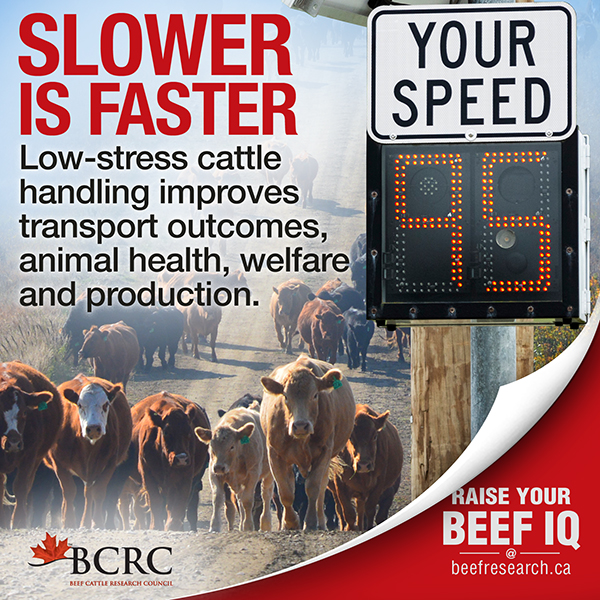
Practicing low-stress animal handling;
Incorporating proper handling facility designs that include adequate traction and comfortable footing;
Applying lime to barn floors following cleaning between fills to make the environmental pH less friendly to infectious lameness-causing bacteria;
Consulting with your veterinarian regarding the potential use of a Fusobacterium necrophorum vaccine to prevent footrot;
Incorporating step-up rations for high-grain diets to reduce the risk of acidosis and laminitis;
Test feeds for potential mycotoxins that may lead to ergot poisoning;
Carefully inspect feet and legs on breeding cattle to ensure they are fit and sound.
3. Griffin, D., Perino, L., and Hudson, D. 1993. G93-1159 Feedlot Lameness. University of Nebraska-Lincoln, Historical Materials from University of Nebraska-Lincoln Extension.
7. Tessitore, E., Schwartzkopf-Genswein, K. S., Cozzi, G., Pajor, E., Goldhawk, C.Brown, F., Janzen, E., Klassen, P.and Dueck, C. 2011. Prevalence of lameness in 3 commercial feedlots in Southern Alberta during summer months. Proceedings of the Canadian Society of Animal Science. pg 75.